 Limited Edition Golden Llama is here! Check out how you can get one.
  Limited Edition Golden Llama is here! Check out how you can get one.
 Offering SPR-BLI Services - Proteins provided for free!
 Get your ComboX free sample to test now!
 Time Limited Offer: Welcome Gift for New Customers !
  Shipping Price Reduction for EU Regions
> Colección de proteínas recombinantes de la vía PD-1
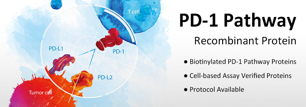
La vía de los puntos del control inmunitario es un aspecto central para la investigación actual sobre el cáncer. LA PD-1 es una de las proteínas del punto de control mejor caracterizada. La unión entre PD-1 y su ligando PD-L1 y/o PD-L2 suprime la activación de las células T y permite a las células cancerosas escapar de la vigilancia inmunitaria del organismo. Este mecanismo sugiere la idea de que la neutralización de la vía PD-1 puede ayudar a reactivar el sistema inmunitario para luchar contra el cáncer.
En los últimos años se han desarrollado múltiples líneas de fármacos anti-PD-1, que han demostrado un enorme potencial en ensayos clínicos recientes. Los pacientes con melanoma, carcinoma de células renales, cáncer de pulmón de células no pequeñas (CPCNP) o cánceres hematológicos responden positivamente a este tratamiento.
ACROBiosystems proporciona una serie completa de productos relacionados con la PD-1 para que los investigadores investiguen y se dirijan a la vía de la PD-1.
ELISA is an assay technique routinely used to characterize protein-protein interaction. It’s often used in initial high-throughput screening (HTS) of compound library. ACROBiosystems has developed a range of pre-biotinylated PD-1 pathway proteins. Biotinylated proteins can serve as two-way antibodies for both capture and detection in ELISA assays. They demonstrate higher specificity and detection sensitivity than traditional antibodies.
| Product Description | Cat. No. | Species | Structure | |
|---|---|---|---|---|
| Pair 1 | Human PD-L1 / B7-H1 Protein, Fc Tag, low endotoxin | PD1-H5258 |  | |
| Pair 2 | Mouse PD-L1 / B7-H1 Protein, Fc Tag | PD1-M5251 |  | |
| Biotinylated Mouse PD-1 / PDCD1, Fc Tag, Avi Tag (AviTagTM) | PD1-M82F4 |  | ||
| Pair 3 | Human PD-1 / PDCD1 Protein, Fc Tag (HPLC-verified), low endotoxin | PD1-H5257 |  | |
| Biotinylated Human PD-L1 / B7-H1, His Tag & Fc Tag, Avi Tag (AviTagTM) | PD1-H82F3 |  | ||
| Pair 4 | Mouse PD-1 / PDCD1 Protein, Fc Tag | PD1-M5259 |  | |
| Biotinylated Mouse PD-L1 / B7-H1, Fc Tag, Avi Tag (AviTagTM) | PD1-M82F5 |  |
To further simplify your research, we’ve developed a PD-1[Biotin]: PD-L1 Inhibitor Screening Kit (Cat. No. EP-101). This assay employs a simple colorimetric ELISA platform, which measures the binding between immobilized human PD-L1 and in-house developed biotinylated PD-1 protein. This product is uniquely suitable for rapid and high-throughput screening of putative PD-1 and PD-L1 inhibitors.
| Molecule | Cat. No. | Product Description | Species | Size |
|---|---|---|---|---|
| PD-1 & PD-L1 | EP-101 | PD-1[Biotin]: PD-L1 Inhibitor Screening Pair | 96 tests, 480 tests |
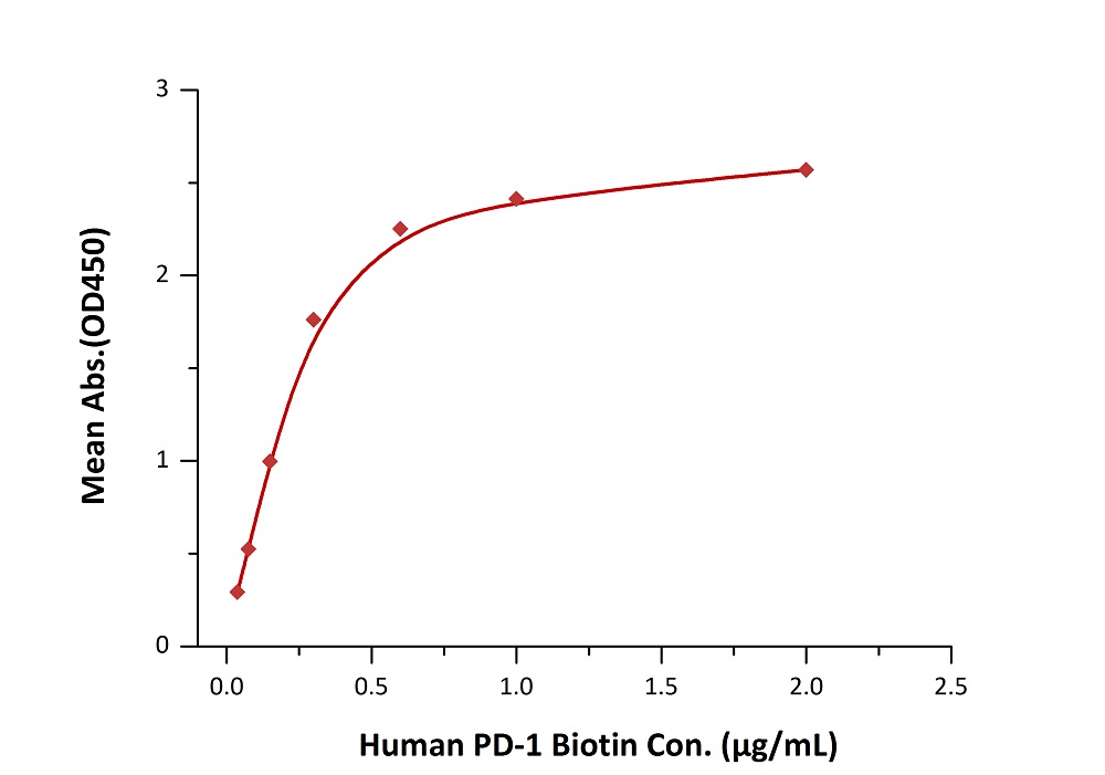
Fig. 2 Immobilized PD-L1 at 2 μg/mL (100 μL/well) can bind biotinylated human PD-1 with a linear range of 0.038 - 0.6 μg/mL when detected by Streptavidin-HRP.
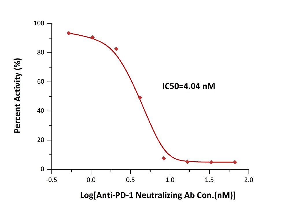
Fig. 3 Inhibition of PD-1-PD-L1 binding by an anti-PD-1 neutralizing antibody is measured by the PD-1 [Biotinylated] : PD-L1 Inhibitor Screening ELISA Assay Pair (Cat. No. EP-101).
Biotinylated proteins can be used along with fluorophore-tagged SA to detect/isolate cells expressing particular surface markers. In a US patent titled "ANTI-PD-1 ANTIBODIES AND METHODS OF USE THEREOF" (US patent 20160159905), the authors (from Pfizer Inc.) used ACROBiosystems' biotinylated PD-L1 (Cat. No. PD1-H82F3) and biotinylated PD-L2 (Cat. No. PD2-H82F6) to compete with candidate antibodies in a binding assay against PD-1 expressing cells. This assay provides measurement for the binding activity of the corresponding antibodies.
Biopanning has gradually become the mainstream approach for antibody development. The AviTagTM biotinylated proteins can be used with SA-coated magnetic beads/surface in biopanning. It’s a much better choice than non-biotinylated, or chemically biotinylated proteins because it offers higher coating density and uniformed antigen presentation.
Please note that the Fc tag is generally undesired for these experiments because it generates false positive results. We have developed AviTagTM PD-1 pathway proteins with only His tag attached. These products are recommended specifically for biopanning.
| Molecule | Cat. No. | Species | Structure | Purity | Features |
|---|---|---|---|---|---|
| PD-1 | PD1-H82E4 |  | |||
| PD-L1 | PD1-H82E5 |  | |||
| PD-L2 | PD2-H82E8 |  |
As a professional supplier of pharmaceutical companies, we have established a cell based assay platform to verify the activity of the target protein, which provide cell based experiment data support for drug screening.
ACROBiosystems used human PD-1 overexpressed 293 cells made in-house in flow cytometry assay and cell based ELISA, and the results showed good performance of PD-L1 (Cat. No. PD1-H5258) and PD-L2 (Cat. No. PD2-H5251) binding to PD-1.
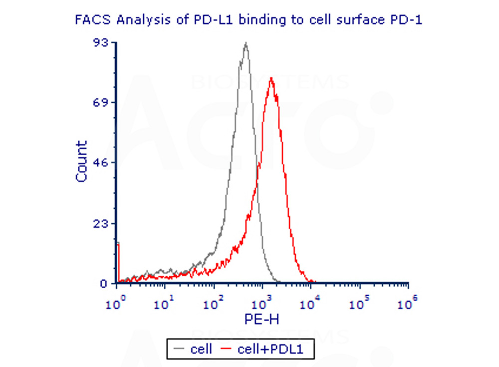
Fig. 4 Flow Cytometry assay shows that recombinant human PD-L1 (Cat. No. PD1-H5258) can bind to 293 cell overexpressing human PD-1.The concentration of PD-L1 used is 10ug/mL
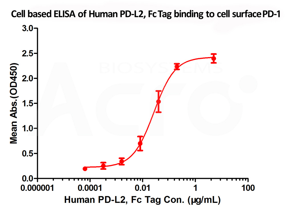
Fig. 5 Immobilized cell surface PD-1 (5x10⁴ of cells per well) can bind Human PD-L2 Protein, Fc Tag (Cat. No. PD2-H5251) with an EC50 of 0.018 μg/mL (Routinely tested).

Fig. 6 FACS analysis shows that the binding of PD-L1 (Cat. No. PD1-H5258) to 293 overexpressing PD-1 was inhibited by increasing concentration of neutralizing anti-hPD-L1 antibody. The concentration of PD-L1 used is 10 μg/mL. The IC50 is 12.92 μg/mL.
1. Hydrogel dual delivered celecoxib and anti-PD-1 synergistically improve antitumor immunity
Authors: Li Y, et al.
Journal: Oncoimmunology
Application: ELISA
Product:PD1-M5228
2. Hererocyclic compounds as immunomodulators
Authors: L Wu, et al.
Journal: US20170107216A1
Application: HTRF Binding Assay
Product: PD1-H5229
3. Hererocyclic compounds as immunomodulators
Authors: J Li, et al.
Journal: US20170145025A1
Application: HTRF Binding Assay
Product: PD1-H5229
4. A novel PD-L1-targeting antagonistic DNA aptamer with antitumor effects.
Authors: Lai WY, et al.
Journal: Mol Ther Nucleic Acids
Application: ELISA-based competition assay
Product: PD1-H5229
5.Anti-pd-1 antibodies and methods of use thereof
Authors: Yasmina Noubia Abdiche, et al.
Journal: US20160159905A1
Application: Flow Cytometry
Product: PD1-H82E5
6. Anti-pd-1 antibodies and methods of use thereof
Authors: Yasmina Noubia Abdiche, et al.
Journal: US20160159905A1
Application: Flow Cytometry
Product: PD2-H82E8
Blank C., et al., 2007, Cancer Immunol. Immunother. 56 (5): 739–45.
He X.-H., et al., 2004, Acta Biochim. Biophys. Sin. 36:284-289.
This web search service is supported by Google Inc.
