Leave message
Can’t find what you’re looking for?
Fill out this form to inquire about our custom protein services!
Inquire about our Custom Services >>


 Limited Edition Golden Llama is here! Check out how you can get one.
  Limited Edition Golden Llama is here! Check out how you can get one.
 Offering SPR-BLI Services - Proteins provided for free!
 Get your ComboX free sample to test now!
 Time Limited Offer: Welcome Gift for New Customers !
  Shipping Price Reduction for EU Regions
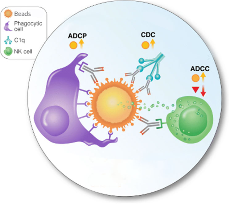
> Insights > Bead-Based Phagocytosis Assay: Cost-effective, Highly Efficient and Convenient to Use With the simple aid of a magnetic field, magnetic beads pre-coupled with biotinylated proteins are well-suited for highly efficient immunocapture, biopanning and virus capture. Apart from these common applications, beads can also be used in antibody-dependent cellular phagocytosis (ADCP) assay to evaluate the phagocytic potential of activated effector cells, an experiment commonly performed in tumor or virus studies.

Fig.1. Antibody-mediated cellular phagocytosis (ADCP) of protein-coupled beads.
A recent study of HIV-1 reported the use of fluorescent bead-based flow cytometric assay to quantify cellular phagocytosis of GP41-coupled beads by monocytes. In the standard bead ADCP assay they performed, protein-coupled fluorescent beads are incubated with antibodies to be adequately opsonized prior to addition to phagocytic cells, and the beads uptak by cells are analyzed by flow cytometry. To further differentiate the surface-attached beads from the intracellular beads, they incorporated a Specific Hybridization Internalization Probe (SHIP) which selectively inactivate the SHIP fluorescence of the attached beads but not of those truly internalized. In this way, the number of beads phagocytosed by single effector cell can also be accurately identified from the fluorescence level. In summary, they reported the method being highly efficient and repeatable.
In bead-based ADCP assay,detection of the phagocytosed beads can be performed with fluorescence technology or electronic microscopy (EM). ACROBiosystems has developed a list of beads products that are suitable to be used in ADCP assays and conveniently visualized with EM. These 2um beads can provide reliable simulation data by mimicking antigen presentation on different tumor cells or virus-infected cells. Additionally, using bead-based assay to measure Fc-mediated phagocytosis has advantages over real cell assays for being inexpensive, convenient to manipulate and can be stored for a long period of time.
Product Lists
| Molecule | Cat. No. | Product Description | Order/Preorder |
|---|---|---|---|
| S protein | MBS-K029 | SARS-CoV-2 Spike Trimer (B.1.1.7) Coupled Magnetic Beads | |
| S protein | MBS-K030 | SARS-CoV-2 Spike Trimer (B.1.351) Coupled Magnetic Beads | |
| S protein | MBS-K031 | SARS-CoV-2 Spike Trimer (P.1) Coupled Magnetic Beads | |
| Spike protein | MBS-K015 | SARS-CoV-2 Spike Trimer-coupled Magnetic Beads | |
| Spike protein | MBS-K016 | SARS-CoV-2 Spike Trimer (D614G)-coupled Magnetic Beads | |
| S protein RBD | MBS-K032 | SARS-CoV-2 Spike RBD (B.1.351) Coupled Magnetic Beads | |
| S protein RBD | MBS-K033 | SARS-CoV-2 Spike RBD (P.1) Coupled Magnetic Beads | |
| S protein RBD | MBS-K034 | SARS-CoV-2 Spike RBD (B.1.1.7) Coupled Magnetic Beads | |
| Spike RBD | MBS-K014 | Human Anti-SARS-CoV-2 Spike RBD Antibody-coupled Magnetic Beads | |
| Spike NTD | MBS-K019 | SARS-CoV-2 Spike NTD-coupled Magnetic Beads | |
| Spike S1 | MBS-K001 | SARS-CoV-2 Spike S1-coupled Magnetic Beads | |
| Spike S1 | MBS-K002 | SARS-CoV-2 Spike RBD-coupled Magnetic Beads | |
| Spike S2 | MBS-K018 | SARS-CoV-2 Spike S2-coupled Magnetic Beads | |
| Nucleocapsid protein | MBS-K017 | SARS-CoV-2 Nucleocapsid Protein-coupled Magnetic Beads | |
| ACE2 | MBS-K013 | Human ACE2-coupled Magnetic Beads | |
| BCMA | MBS-K004 | Human BCMA-coupled Magnetic Beads | |
| CD19 | MBS-K005 | Human CD19-coupled Magnetic Beads | |
| CD3 epsilon | MBS-K026 | Human CD3E-coupled Magnetic Beads | |
| CD38 | MBC-K010 | Human CD38-coupled Magnetic Beads | |
| CD3E & CD3D | MBS-K003 | Human CD3E & CD3D Heterodimer-coupled Magnetic Beads | |
| CD73 | MBS-K022 | Human CD73-coupled Magnetic Beads | |
| EGF R | MBE-K012 | Human EGFR-coupled Magnetic Beads | |
| EGFRvIII | MBS-K020 | Human EGFRvIII-coupled Magnetic Beads | |
| Her2 | MBS-K006 | Human HER2-coupled Magnetic Beads | |
| HGF R | MBS-K021 | Human HGF R-coupled Magnetic Beads | |
| PSMA | MBP-K007 | Human PSMA-coupled Magnetic Beads | |
| ROR1 | MBR-K009 | Human ROR1-coupled Magnetic Beads | |
| Siglec-3 | MBC-K008 | Human CD33-coupled Magnetic Beads | |
| Streptavidin | SMB-B01 | Magnetic Beads™ Streptavidin |
Reference:
1.Duchemin M, Tudor D, Cottignies-Calamarte A, Bomsel M. Antibody-Dependent Cellular Phagocytosis of HIV-1-Infected Cells Is Efficiently Triggered by IgA Targeting HIV-1 Envelope Subunit gp41. Front Immunol. 2020 Jun 9;11:1141. doi: 10.3389. Retrieved from https://www.frontiersin.org/articles/10.3389/fimmu.2020.01141/full
2.Lloyd, Y.M., Ngati, E.P., Salanti, A. et al. A versatile, high through-put, bead-based phagocytosis assay for Plasmodium falciparum . Sci Rep 7, 14705 (2017). Retrieved from https://doi.org/10.1038/s41598-017-13900-4
This web search service is supported by Google Inc.


