Jul 13, 2026
 Page Views:1602


In vivo CAR-T is rapidly emerging as one of the most closely watched frontier tracks in cell therapy. Recently, positive clinical updates from Kelonia, Legend Biotech, and others have rekindled market expectations for this "next-generation CAR-T" technology. Unlike conventional CAR-T, which requires complex, multi-step processes—including leukapheresis, ex vivo genetic modification, and reinfusion—in vivo CAR-T leverages delivery systems to generate CAR-T cells directly within the patient's body, offering the potential for lower costs, shorter treatment cycles, and broader patient access. Among the enabling platforms, targeted lipid nanoparticles (tLNPs) stand out as one of the most promising, owing to their efficient and safe nucleic acid delivery capabilities. tLNPs can precisely deliver CAR-encoding mRNA to T cells, enabling in situ CAR-T cell generation. This strategy has already shown encouraging therapeutic potential across oncology, autoimmune diseases, and cardiovascular disorders. However, despite the development of various tLNP systems targeting distinct T-cell surface receptors, substantial differences in delivery efficiency and in vivo CAR-T generation persist across targets—and the underlying mechanisms remain incompletely understood.
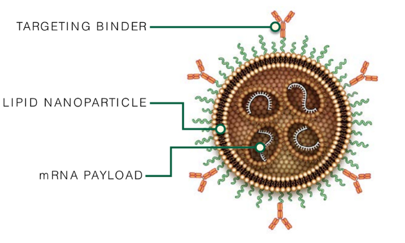
Schematic of targeted lipid nanoparticles (tLNPs) (Source: Science (New York, N.Y.) vol. 388,6753 (2025): 1311-1317.)
tLNP Endocytosis Efficiency: The Core Driver of In Vivo CAR-T Generation
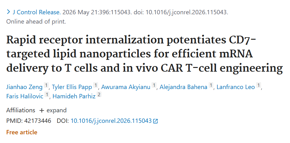
Recently, a landmark study from the team led by Hamideh Parhiz, co-founder of Capstan Therapeutics, systematically evaluated—for the first time—how different T-cell targeting receptors influence tLNP delivery efficiency. The findings upend conventional assumptions: receptor expression abundance is not the key determinant of delivery performance. Instead, receptor internalization efficiency emerges as the dominant factor governing mRNA delivery and subsequent in vivo CAR-T generation. This insight provides a fresh paradigm for designing next-generation in vivo CAR-T delivery vehicles.
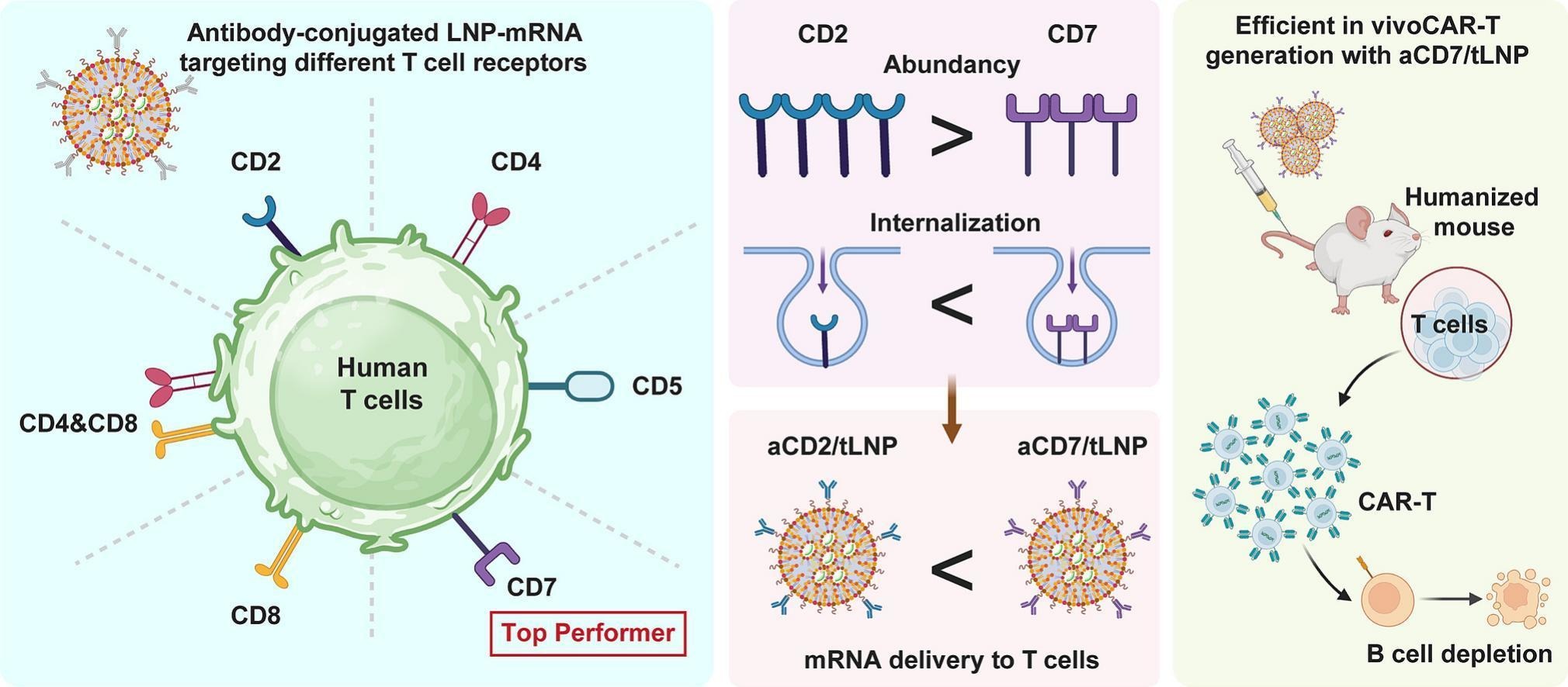
The study systematically compared mRNA delivery by tLNPs targeting CD2, CD4, CD5, CD7, CD8, and CD4/CD8 dual-targeting constructs in primary human T cells, and identified the critical factors driving delivery efficacy:
(1) High abundance does not guarantee high efficiency:
Both transcriptomic and flow cytometric analyses confirmed that CD2 is the most highly expressed receptor on T-cell surfaces—its protein levels exceed those of CD7 by >2-fold and CD5 by >3-fold. According to the traditional "abundance-determines-efficacy" hypothesis, CD2-targeted tLNPs should have outperformed all others. Yet the results showed the opposite: aCD2/tLNP ranked only third in delivery efficiency, with mean fluorescence intensity (MFI) just one-fifth that of aCD7/tLNP. In other words, high T-cell receptor abundance does not ensure superior transfection efficiency for tLNPs targeting that receptor.
(2) Internalization kinetics is the decisive metric:
To probe the mechanistic basis for these differences, the researchers employed a pH-sensitive FabFluor labeling system to monitor real-time internalization of receptor–antibody complexes. Strikingly, the aCD7 antibody exhibited the highest internalization level—surpassing even CD71 (transferrin receptor, a classic high-endocytosis positive control) within the 4‑hour observation window—whereas the aCD2 signal remained extremely weak.
(3) Intracellular mRNA accumulation is strongly correlated with internalization:
Using Cy5‑labeled mRNA tracing, the study demonstrated that intracellular mRNA accumulation correlates highly with antibody–receptor internalization levels (Pearson r = 0.894, p < 0.0001). The aCD7/tLNP group achieved the highest intracellular mRNA signal within just 4 hours.
(4) High-internalization aCD7/tLNP enables potent in vivo CAR-T cell generation:
In humanized mouse models, aCD7/tLNP loaded with anti‑CD20 CAR mRNA—at a low dose of 2.5 μg per mouse—successfully generated CAR-T cells in the spleen within 24 hours (approximately 25% CAR expression in CD4⁺ T cells and 40% in CD8⁺ T cells), driving near-complete B‑cell depletion. This efficiency significantly outperformed the aCD2/tLNP control under the same conditions, with serum biochemistry assays indicating a favorable safety profile.
Conclusion
This study provides the first systematic evidence that, for T‑cell‑targeted delivery, receptor internalization efficiency—not receptor expression abundance—is the pivotal factor governing mRNA delivery and in vivo CAR‑T generation. For companies developing in vivo CAR‑T delivery systems, these findings underscore the importance of incorporating receptor internalization capacity as a key evaluation metric. Establishing robust assays to monitor internalization can provide a more scientifically grounded basis for selecting delivery targets and optimizing vector designs.
Moreover, assessing receptor internalization offers a significant front‑loaded R&D advantage: rather than waiting for CAR expression data or even in vivo efficacy results to validate delivery strategies, internalization measurements can identify potential bottlenecks early, flag low‑efficiency targets or delivery approaches, reduce trial‑and‑error costs, shorten development timelines, and accelerate iterative optimization. In the increasingly competitive in vivo CAR‑T landscape, advancing key evaluation metrics upstream may become a critical strategy for enhancing R&D productivity and success rates. As the field continues to evolve rapidly, further elucidation of delivery mechanisms will uncover additional determinants, driving continued efficiency gains and expediting the transition of in vivo CAR‑T from clinical innovation to broad industrial application.
【New Product Launch】DTrack™ Endocytosis Efficiency Detection Kit
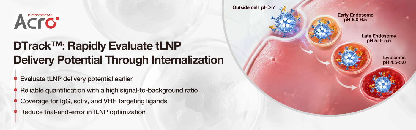
With the rapid advancement of in vivo CAR‑T delivery technologies, scFvs and VHHs—owing to their small molecular size, low steric hindrance, and reduced immunogenicity—have become the predominant antibody formats for tLNP surface targeting. To address the pressing need for efficient, user‑friendly tools to assess tLNP endocytosis during in vivo CAR‑T development, ACROBiosystems has launched the DTrack™ series of pH‑sensitive dye‑labeled universal antibodies. This portfolio is compatible with various antibody formats—including IgG, scFv, and VHH—and enables rapid endocytosis evaluation, empowering researchers to assess delivery potential, screen optimal targeting ligands, and provide practical evidence for achieving efficient in vivo CAR‑T engineering.
Assay Principle
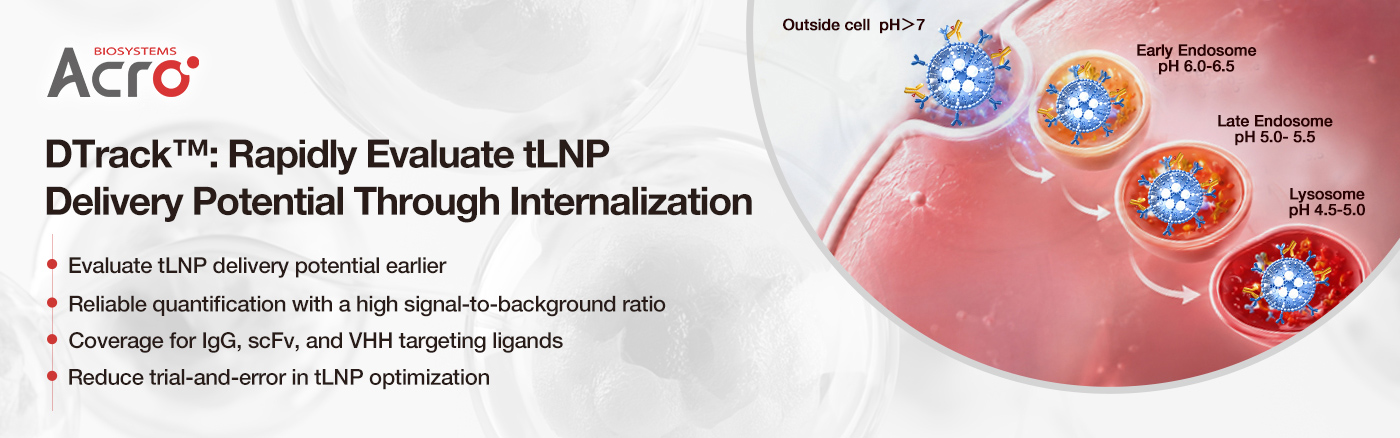
This kit series employs pH‑sensitive dye labeling technology, which exhibits minimal fluorescence in the extracellular neutral environment (pH ≈ 7.4). Upon binding to targeting antibodies on the LNP surface and subsequent receptor‑mediated internalization into acidic endosomal/lysosomal compartments (pH 4.5–5.5), the pH‑sensitive dye is activated and emits a bright red fluorescence. By monitoring fluorescence intensity and the percentage of positive cells in real time, researchers can rapidly and quantitatively evaluate the endocytosis efficiency mediated by different targeting antibodies, enabling swift identification of optimal ligand candidates.
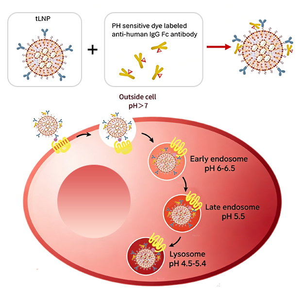
Product Advantages
- Comprehensive Coverage: Offers a complete endocytosis detection system for mainstream tLNP targeting ligands, including IgG, scFv, and VHH formats.
- Accurate and Reliable Detection: The "off–on" fluorescence design of the pH‑sensitive dye ensures low background and high signal‑to‑noise ratio, precisely reflecting receptor internalization dynamics.
- Accelerated R&D Efficiency: Streamlines screening of high‑potential ligands and optimization of delivery systems.
Validation Data
pH-sensitive dye-labeled Anti-G4S linker antibody binds to the Anti-CD8 antibody modified on the surface of LNP containing a G4S linker. Upon binding to CD8 receptors on T cells, the complex is internalized, and red fluorescence can be detected in the acidic intracellular environment, thereby allowing assessment of the internalization efficiency of tLNP.
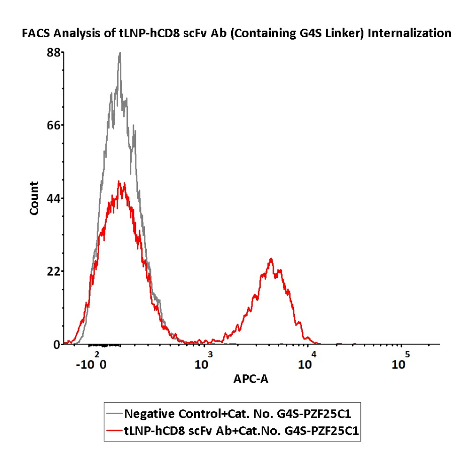
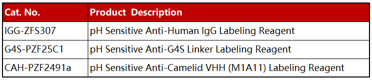
tLNP-hCD8 scFv Ab (containing G4S linker) and negative control tLNP were labeled with pH Sensitive Anti-G4S Linker Labeling Reagent (Cat. No. G4S-PZF25C1). CD3+ T cells were separately treated with pH-sensitive Dye-Labeled tLNP-hCD8 scFv Ab conjugates or negative control for 4 hours, then analysis by Flow cytometric. APC signal was used to evaluate the activity.
Request ProtocolpH-sensitive dye-labeled Anti-VHH antibody binds to the Anti-CD8 VHH antibody modified on the surface of LNP. After binding to CD8 receptors on T cells and undergoing internalization, red fluorescence is detectable in the acidic intracellular environment, enabling evaluation of tLNP uptake efficiency.
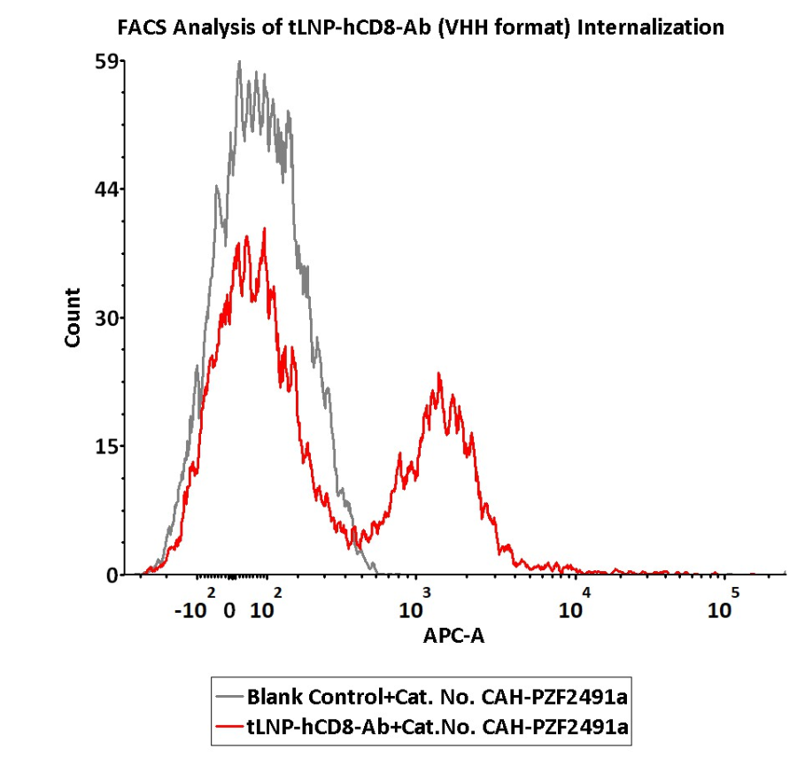
tLNP-hCD8-Ab (VHH format) were labeled with pH Sensitive Anti-Camelid VHH (M1A11) Labeling Reagent (Cat. No. CAH-PZF2491a). CD3+ T cells were separately treated with pH-sensitive Dye-Labeled tLNP-hCD8 VHH Ab conjugates or pH-sensitive Dye alone for 4 hours, then analysis by Flow cytometric. APC signal was used to evaluate the activity.
Request Protocol• pH Sensitive Anti-Human IgG Labeling Reagent detects internalization of LNP conjugated with CD8 antibody (human Fc). Upon binding to the antibody on the LNP surface and internalization into acidic endosomes or lysosomes, red fluorescence can be detected.
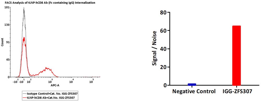
tLNP-hCD8 Ab and LNP-Human IgG1 Kappa Isotype Control were labeled with pH Sensitive Anti-Human IgG Labeling Reagent (Cat. No. IGG-ZFS307). CD3+ T cells were separately treated with pH-sensitive Dye-Labeled tLNP-hCD8 Ab or isotype control conjugates for 4 hours, then analysis by Flow cytometric. APC signal was used to evaluate the activity.
Request ProtocolRecommended Additional Products
Delivery Vector Surface Antibody Density Characterization Tools
Universal CAR‑Positive Rate Detection Tools
Anti‑Idiotype Antibodies (FMC63, C11D5.3)

FAQ
Q1: What is a tLNP endocytosis assay?
A: A tLNP endocytosis assay measures how efficiently targeted lipid nanoparticles (tLNPs) are internalized by target cells after binding to specific surface receptors. Using pH-sensitive fluorescent probes, these assays enable rapid, quantitative evaluation of receptor-mediated uptake, helping researchers optimize ligand selection and delivery performance for in vivo CAR-T and other nucleic acid therapies.
Q2: Why is receptor internalization important for in vivo CAR-T delivery?
A: Research has revealed that, contrary to conventional wisdom, the expression abundance of target receptors is not the key determinant of delivery efficacy. Instead, receptor internalization efficiency is the critical factor governing mRNA delivery and in vivo CAR‑T generation. This finding suggests that companies developing in vivo CAR‑T delivery systems should incorporate receptor internalization capacity as a key evaluation metric. By establishing robust assays to detect and monitor internalization, they can provide a more scientifically grounded basis for selecting delivery targets and optimizing vector designs.
Q3: How do pH-sensitive endocytosis assays work?
A: These assays use pH‑sensitive dyes that are almost non‑fluorescent at neutral extracellular pH. When the dye is attached to targeting antibodies on LNPs, the particles are taken up via receptor‑mediated endocytosis into acidic endosomes/lysosomes. There, the dye is activated and emits bright red fluorescence. By measuring the fluorescence intensity and the percentage of positive cells, you can directly compare how efficiently different targeting ligands drive internalization—helping you quickly identify the best candidate.
Q4: How can endocytosis assays accelerate tLNP development?
A:By measuring receptor internalization early in the development process, endocytosis assays help researchers identify high-performing targeting ligands, optimize nanoparticle formulations, reduce trial-and-error experimentation, and improve the efficiency of mRNA delivery before advancing to in vivo studies.
Reference
Zeng J, Papp TE, Akyianu A, et al. Rapid receptor internalization potentiates CD7‑targeted lipid nanoparticles for efficient mRNA delivery to T cells and in vivo CAR T‑cell engineering. Preprint. bioRxiv. 2026;2026.01.23.701374. Published 2026 Jan 26. doi:10.64898/2026.01.23.701374