



Microelectrode Array (MEA) Service
Cardiotoxicity remains a leading cause of candidate drug attrition and clinical termination, often manifesting as arrhythmias, electrophysiological abnormalities, and impaired contractile function. Conventional 2D cell models fail to recapitulate the complex tissue architecture and functionality of the human heart, while animal models exhibit significant interspecies variability. Moreover, reliable, real-time functional assays are essential to capture drug-induced electrophysiological and contractile changes.
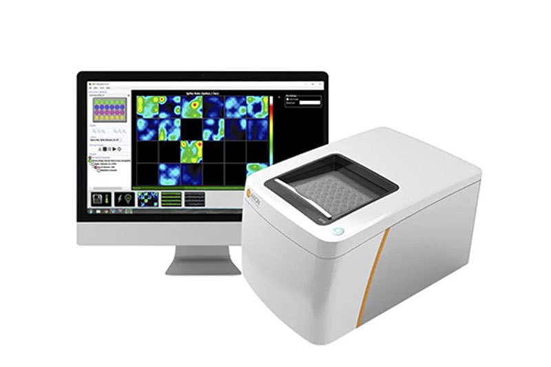
Leveraging our proprietary human iPSC-derived cardiac organoids, together with microelectrode array (MEA)-based real-time electrophysiological recording and complementary functional assays, we have established an integrated Cardiac Organoid MEA Cardiotoxicity Assessment Service. This platform enables comprehensive evaluation of drug-induced changes in cardiac function, rhythm stability, and proarrhythmic risk, providing reliable data to support early-stage safety screening, candidate prioritization, preclinical cardiotoxicity assessment, and informed decision-making during drug development.
Service Advantages
| Core Advantages | Description |
|---|---|
| Integrated Organoid-MEA Platform | Self-developed human iPSC-derived cardiac organoids are integrated with MEA electrophysiology to support one-stop cardiac safety assessment. |
| Human-Relevant 3D Cardiac Model | Cardiac organoids display spontaneous beating activity and tissue-like functional features, helping improve the translational relevance of in vitro cardiotoxicity evaluation. |
| Real-Time Functional Detection | MEA recording enables non-invasive monitoring of drug-induced changes in electrical activity, rhythm stability, and repolarization-related responses. |
| Flexible Study Design | The service supports baseline recording, drug treatment, concentration-response testing, and customized project designs. |
Standard Workflow & Service Scope
| Service Module | Key Description | Readouts |
|---|---|---|
| 1. Cardiac Organoid Preparation | Human iPSC-derived cardiac organoids are prepared through ACROBiosystems’ standardized differentiation and culture workflow. | Spontaneously beating cardiac organoids ready for MEA assay |
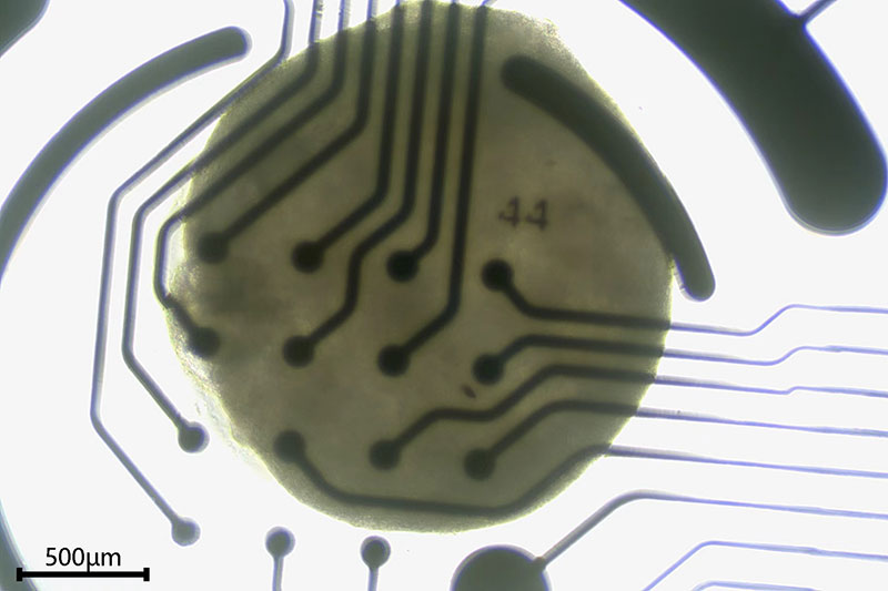
| 2. MEA Plate Seeding & Stabilization | Cardiac organoids are seeded onto MEA electrodes and stabilized before testing. | Stable baseline beating and electrophysiological activity |
| 3. Baseline MEA Recording | Baseline electrophysiological profiles are recorded before compound treatment. | Baseline beat rate, FPD/FPDc, spike amplitude, rhythm stability |
| 4. Drug Treatment & Dose Design | Acute exposure and concentration-gradient designs can be customized based on project objectives. Optional repeated dosing or recovery assessment can be discussed based on feasibility. | Compound-induced functional response across dose groups |
| 5. Real-Time MEA Functional Recording | MEA is used to monitor drug-induced changes in cardiac electrical activity. | Field potential waveform, beat rate, FPD/FPDc, spike amplitude, rhythm irregularity / arrhythmia-like events |
| 6. Data Analysis & Report Delivery | Electrophysiological responses are analyzed and summarized in a project report. | Cardiac functional risk assessment and project report |
| (Optional) Customized Add-on Readouts | Additional assays can be included based on project needs and feasibility. | Calcium Imaging: Assessment of calcium transient and excitation-contraction-related function. |
| cTnT Immunostaining: Evaluation of cardiac structural marker expression. | ||
| qPCR: Analysis of cardiac stress, ion channel, and mechanism-related gene expression. | ||
| ELISA: Detection of secreted biomarkers or functional response markers. | ||
| Viability Assay: Assessment of cell viability and cytotoxicity. | ||
| Customized Analysis: Project-specific readout design. |
Service Case
Electrophysiological profiling of hiPSC-derived cardiac organoids by MEA
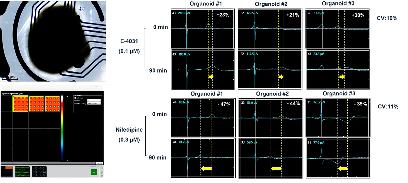
MEA recordings showed stable electrical activity across multiple electrodes in cardiac organoids. Following treatment with E-4031, a hERG/IKr potassium channel blocker, and nifedipine, an L-type calcium channel blocker, the organoids showed compound-related changes in MEA-derived electrophysiological readouts, supporting the potential use of this model for cardiac functional and safety assessment.
- Introduction
- Service Advantages
- Standard Workflow & Service Scope
- Service Case