CD8 in Immunotherapy: CAR-T Engineering, in vivo CAR, and CD8-LNP Applications
 Publication Date:2026-06-01
 Page Views:633
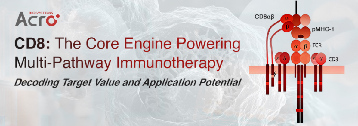
Within the finely tuned regulatory network of the immune system, the CD8 co-receptor plays an irreplaceable central role. As a key molecule on the surface of CD8+ T cells, CD8 not only participates in antigen recognition and T cell activation but has also emerged as a critical target and tool for next-generation immunotherapies.
Molecular Architecture and Domains of CD8
CD8 exists as a dimer, primarily in two isoforms: CD8αα homodimer and CD8αβ heterodimer. These two forms differ significantly in tissue distribution, ligand-binding characteristics, and signaling capacity.
Structurally, CD8 comprises four functional modules: the extracellular domain adopts an immunoglobulin-like fold that directly interacts with the α3 domain of MHC-I molecules, stabilizing the TCR/pMHC complex; the hinge region is rich in proline, threonine, and serine, exhibiting high conformational flexibility, with O-glycosylation patterns that dynamically change with T cell developmental stages, influencing binding affinity and signal transduction; the transmembrane domain is a type I single-pass α-helix, stabilized as a dimer via conserved cysteine residues forming disulfide bonds, determining membrane localization and stability; in the cytoplasmic tail, CD8α recruits Lck kinase through a CxC motif forming a zinc-ion coordination complex to initiate signaling, whereas CD8β undergoes palmitoylation-mediated lipid raft enrichment to enhance signaling efficiency, while the cytoplasmic tail also engages in competitive binding with LAT, participating in fine-tuning signal strength.
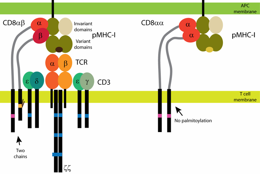
Figure 1.CD8 co-receptor (source: Front Immunol.)
The overall conformations of CD8αα and CD8αβ are highly conserved (Cα RMSD ~1.16 Å), yet the dimer interface areas differ, suggesting subtle distinctions in ligand-binding modes. This refined structural division of labor enables CD8 to perform multiple functions in antigen recognition, signal transduction, and immune response regulation, and has also made it an important target and tool for innovative therapies such as antibody drugs and in vivo CAR-T.
Functional Mechanisms of CD8
• Synergy in Antigen Recognition
For low-affinity TCRs (KD > 30 μM), CD8 significantly enhances the stability of the TCR/pMHC complex by binding the MHC-I α3 domain, boosting antigen recognition sensitivity. For high-affinity TCRs (KD < 10 μM), CD8 engagement is non-essential. This mechanism defines the "threshold-tuning" role of CD8 in immune responses.
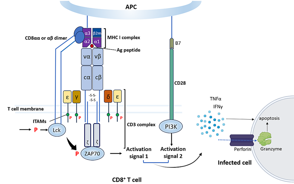
Figure 2.CD8+ T cell activation (source: Biochem Pharmacol.)
• Initiation and Amplification of Signal Transduction
CD8 recruits Lck via its cytoplasmic tail; Lck then phosphorylates the ITAMs of the CD3 complex, initiating downstream ZAP-70, LAT, and SLP-76 cascades, ultimately inducing T cell activation, proliferation, and effector functions. CD8αβ is superior to CD8αα in signal amplification efficiency, primarily attributable to CD8β-mediated lipid raft localization and Lck enrichment.
• Cell Development and Subset Specificity
CD8αβ participates in positive selection and maturation of CD8+ T cells in the thymus, whereas CD8αα mainly exerts a co-inhibitory role in intestinal intraepithelial lymphocytes, regulating immune tolerance. Regarding cell subset distribution, CD8αβ is primarily found on conventional cytotoxic T cells, memory T cells, NKT cells, MAIT cells, and γδ T cells, serving as a co-receptor to enhance T cell affinity for pMHC. CD8αα is distributed on intestinal epithelial T cells, dendritic cells, some memory T cells, and NK cells, with CD8αα functioning in immune homeostasis regulation.
• Interaction Spectrum with MHC-I
The binding affinity of CD8 for the MHC-I α3 domain typically falls in the KD range of 10–500 μM. Notably, certain MHC-I alleles (e.g., HLA-A*68:01, HLA-E) bind CD8 extremely weakly and are defined as "non-binders." Additionally, CD8 interacts with non-classical MHC-I molecules such as MR1, TL, and CD1, expanding its functional boundaries in innate and adaptive immunity.
Hot Development Strategies for CD8-Targeting Drugs
1. Therapeutic Antibodies: Three Pathways for Functional Modulation
Based on functional phenotypes, anti-CD8 monoclonal antibodies can be classified into three categories:
- Blocking Antibodies: Inhibit T cell activation through steric hindrance or by altering immune synapse conformation; primarily applied in transplantation tolerance and autoimmune disease models.
- Enhancing Antibodies: Stabilize the CD8/MHC-I complex or promote signaling to augment T cell effector functions, possessing potential for anti-tumor applications.
- Neutral Antibodies: Used for immuno-PET imaging without interfering with T cell function, enabling non-invasive monitoring of CD8+ T cell distribution in vivo.
Building on monoclonal antibodies, next-generation multispecific antibodies further enhance therapeutic precision and synergistic effects by simultaneously targeting CD8 and tumor antigens or immunomodulatory molecules. Representative clinical-stage drugs include AstraZeneca’s AZD5492 (a trispecific antibody) and AB248 (an antibody fusion protein) from Asher Bio in collaboration with AstraZeneca, both currently in clinical development.
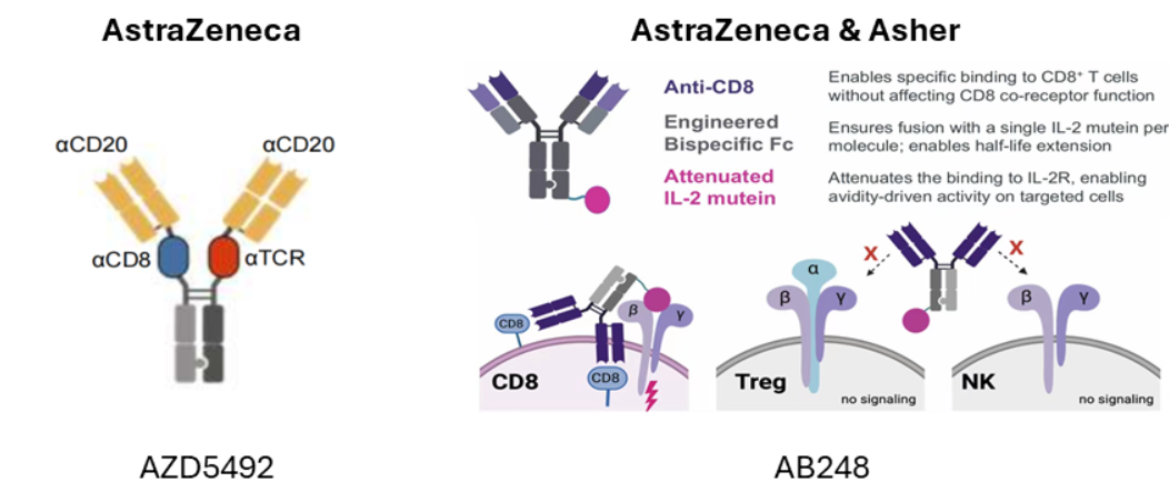
Figure 3.Mechanism of action of AZD5492 and AB248 (source: SITC Annual Meeting)
2. In Vivo CAR: CD8 as a Targeting Element
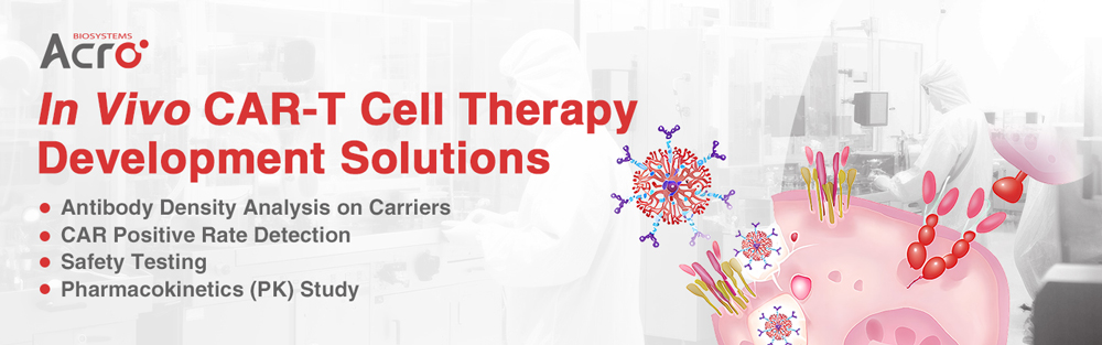
In vivo CAR-T involves intravenous administration of a delivery vector that genetically reprograms T cells directly inside the patient’s body to express chimeric antigen receptors (CARs), thereby transiently or permanently transforming them into CAR-T cells capable of precisely recognizing and eliminating target cells. Often, delivery vehicles such as lipid nanoparticles (LNPs) or lentivirus (LV) are surface-conjugated with targeting antibodies (e.g., CD8 antibody), forming antibody-conjugated delivery vectors that significantly enhance their recognition and localization to specific cells like T cells, enabling precision therapy.
A representative advance in this strategy comes from Capstan Therapeutics and the University of Pennsylvania team, published in Science in 2025, which demonstrated for the first time the feasibility of directly generating functional CAR-T cells in vivo in non-human primates. The team designed a novel ionizable lipid, L829, and modified it with a CD8 antibody, achieving highly selective mRNA delivery to CD8+ T cells while avoiding non-specific activation of CD4+ T cells.
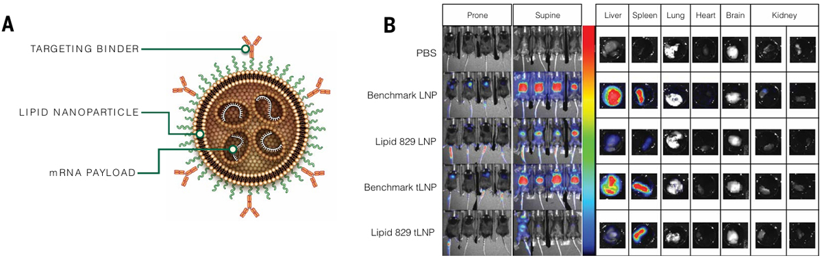
Figure 4.Schematic of tLNP (source: Science)
3. Structural Elements in Cell Therapy: Engineering Applications of CD8 Hinge and Transmembrane Domains
In CAR-T cell therapy, CD8 is mainly utilized as a potency-enhancing element rather than an independent target. The hinge and transmembrane domains of CD8α are widely used in CAR construct design, influencing CAR flexibility, expression levels, signal strength, and resistance to activation-induced cell death.
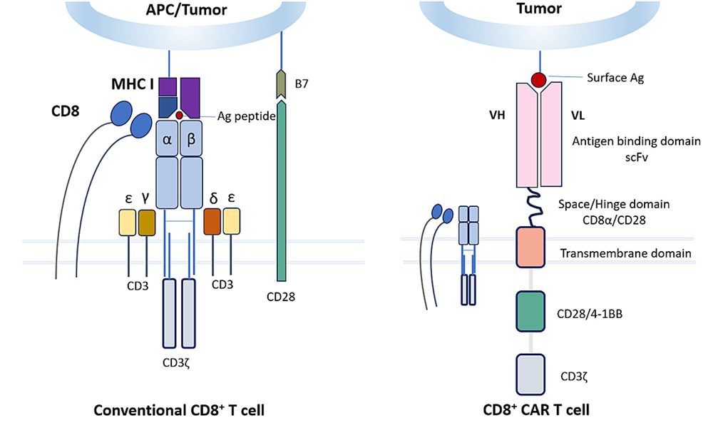
Figure 5.Differences between conventional CD8+ T cells and CAR-T cells (source: Biochem Pharmacol.)
Overview of Clinical-Stage CD8-Based Pipelines (Partial)
From the engineering application of CD8 hinge and transmembrane domains in CAR constructs to CD8 antibody-modified in vivo CAR-T delivery, multiple CD8-based strategies have spawned numerous clinical-stage drug candidates. The table below summarizes a selection of representative pipelines currently in Phase I/II trials.
Table 1. Clinical pipeline overview (source: Pharmacodia)
| Drug Name | Global Highest Development Status & Date | Originator | Active Company | Therapeutic Area / Indication | Drug Type |
|---|---|---|---|---|---|
| 89Zr-Df-crefmirlimab | Phase III 2025-02-01 | Imaginab Inc; | DynamiCure Biotechnology Ltd; Imaginab Inc; National Cancer Institute; National Institute Of Neurological Disorders And Stroke; Pfizer; University Medical Center Groningen; Yantai Dongcheng Biochemicals Co.,Ltd. | Non-Small Cell Lung Cancer; Solid Tumors; Merkel Cell Carcinoma; Melanoma; Renal Cell Carcinoma; Multiple Sclerosis; Progressive Multifocal Leukoencephalopathy; Lymphoma; Cutaneous Melanoma; | Humanized Monoclonal Antibody; Immunoglobulin; In Vivo Diagnostic Agent; Antibody Fragment; Radiolabeled Antibody; Diagnostic Radiopharmaceutical; 89Zr; Radionuclide Drug Conjugate; |
| PDS-0101 | Phase III 2025-05-30 | Pds Biotechnology Corp; | Farmacore; National Cancer Institute; Pds Biotechnology Corp; University of Texas MD Anderson Cancer Center; Weill Medical College of Cornell University; | Oropharyngeal Cancer; Squamous Cell Carcinoma; Head and Neck Cancer; HIV Infection; Anal Cancer; Cervical Cancer; Human Papillomavirus Infection; Vulvar Cancer; | Therapeutic Vaccine; Subunit Vaccine; |
| LB-4330 | Phase III 2025-10-01 | Shanghai Jianxin Biomedical Technology Co., Ltd. | Shanghai Jianxin Biomedical Technology Co., Ltd.;Ningbo Jianxin Biomedical Technology Co., Ltd.; | Cervical Cancer; Solid Tumors; | Bispecific Antibody; |
| MCPyV TAg-specific polyclonal autologous CD8-positive T-cell immunotherapy | Phase II | Fred Hutchinson Cancer Research Center; National Cancer Institute; | Fred Hutchinson Cancer Research Center; National Cancer Institute; | Merkel Cell Carcinoma; | T Cell Therapy; |
| Autologous anti-NY-ESO1 T-cell receptor gene-engineered peripheral blood lymphocytes (Roswell) | Phase II | Roswell Park Cancer Institute; | Roswell Park Cancer Institute; | Tumor; | TCR Cell Therapy; Gene Therapy; |
| Anti-CD30 CAR T-cell therapy | Phase II | Chinese PLA General Hospital; | Cellular Biomedicine Group (Shanghai) Co., Ltd..; Chinese PLA General Hospital; | Hodgkin Disease; | Chimeric Antigen Receptor T Cell Therapy (CAR-T); Gene Therapy; |
| 4-1BB CTL adoptive T cell therapy (Eutilex) | Phase II | Eutilex; | Eutilex; | Tumor; | T Cell Therapy; |
| Nelipepimut S | Phase II 2014-10-01 | University of Texas MD Anderson Cancer Center; Uniformed Services University Of The Health Sciences; Henry M. Jackson Foundation For The Advancement Of Military Medicine; | Cancer Insight Llc; | Breast Cancer; | Therapeutic Vaccine; |
| GEN-009 | Phase II 2018-08-29 | Genocea Biosciences Inc; | Genocea Biosciences Inc; | Non-Small Cell Lung Cancer; Urothelial Carcinoma; Cutaneous Melanoma; Renal Cell Carcinoma; Head and Neck Squamous Cell Carcinoma; | Therapeutic Vaccine; |
| EBViNT Cell (Eutilex) | Phase II 2018-12-14 | Eutilex; | Eutilex; | Extranodal NK-T-Cell Lymphoma; | T Cell Therapy; |
| ZED88082A | Phase II 2019-02-14 | Umcg The University Medical Center Groningen; | University Medical Center Groningen; | Metastatic Cancer; | Monoclonal Antibody; |
| IMA-203 CD8 | Phase II 2019-05-14 | Immatics Us Inc; | Immatics Us Inc; | Metastatic Cancer; | TCR Cell Therapy; |
| VE-800 | Phase II 2020-01-23 | Vedanta Biosciences Inc; | Vedanta Biosciences Inc; | Melanoma; Colorectal Cancer; Gastroesophageal Junction Tumor; Gastric Cancer; Metastatic Cancer; | Live Biotherapeutic Product; |
| CD8+ memory T cell therapy (Stanford University) | Phase II 2020-02-27 | Stanford University; | National Cancer Institute; National Institutes of Health; | Leukemia; Myelodysplastic Syndrome; Acute Lymphoblastic Leukemia; Acute Myeloid Leukemia; Chronic Myeloid Leukemia; | T Cell Therapy; |
| MAGE-A1-specific T cell receptor-transduced autologous T-cell therapy | Phase II 2020-11-13 | SignalOne Bio Inc; | Fred Hutchinson Cancer Research Center; | Non-Small Cell Lung Cancer; Lung Cancer; Urothelial Carcinoma; Breast Cancer; Triple-Negative Breast Cancer; Solid Tumors; | Gene Therapy; TCR Cell Therapy; |
| RB-0003 | Phase II 2021-05-19 | Biontech Se; Tron; | Biontech Se; | Melanoma; | Therapeutic Vaccine; RNA Vaccine; |
| EU-101 | Phase II 2021-05-31 | Eutilex; | Eutilex; Zhejiang Huahai Pharmaceutical Co., Ltd. | Non-Small Cell Lung Cancer; Prostate Cancer; Renal Cell Carcinoma; Solid Tumors; | Humanized Monoclonal Antibody; |
| Insulin growth factor receptor type 1 antisense therapy (Imvax) | Phase II 2023-03-20 | Thomas Jefferson University; | Imvax; | Glioblastoma; | Immune Cell Therapy; |
| SNA-006 | Phase II 2024-04-01 | Suzhou Smartnuclide. Co. Ltd | The First Hospital of China Medical University; Suzhou Smartnuclide. Co. Ltd | Tumor; Solid Tumors; | Nanobody; Radiolabeled Antibody; In Vivo Diagnostic Agent; 68Ga; Diagnostic Radiopharmaceutical; Radionuclide Drug Conjugate; |
| AZD-5492 | Phase II 2024-09-18 | AstraZeneca; | AstraZeneca Limited; AstraZeneca Global R&D (China) Co., Ltd.; AstraZeneca Nijmegen BV; AstraZeneca; | B-Lymphocytic Leukemia; B-Cell Lymphoma; Myositis; Systemic Lupus Erythematosus; Rheumatoid Arthritis; | Trispecific Antibody; |
High-Quality Tool Proteins: Empowering CD8-Related Drug Development
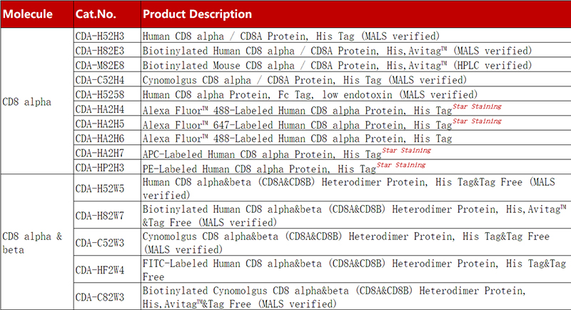
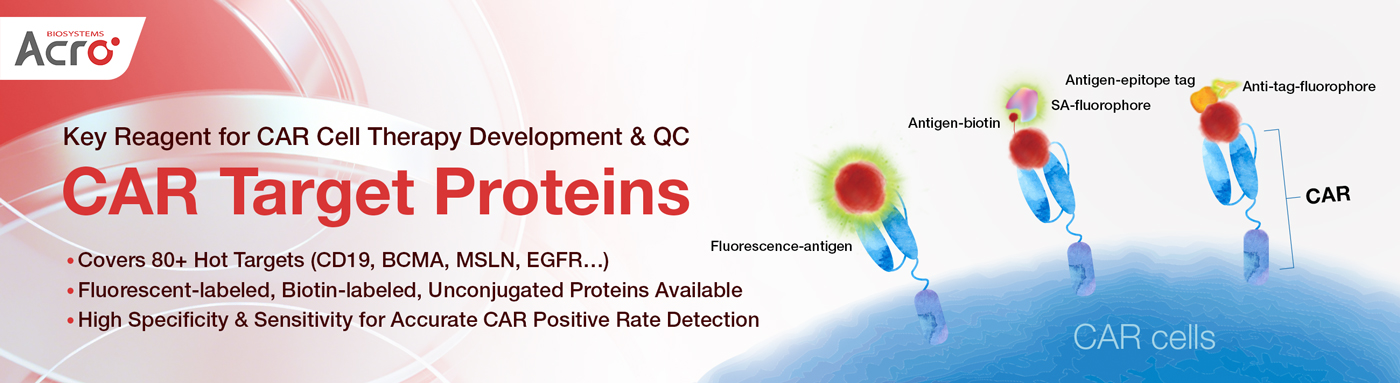
In early screening, functional validation, and quality control of CD8-targeting drugs, high-quality recombinant proteins serve as indispensable core tools. ACROBiosystems provides biotin-labeled, fluorescent-labeled, and unconjugated forms of CD8α and CD8αβ proteins, which are widely applicable in immunoassays, antibody screening, activity verification, and measurement of antibody density on in vivo CAR delivery vehicles.
Product Advantages:
- High Purity and Homogeneity: SEC-MALS verified purity ≥95%, uniform molecular weight, free of aggregates or degradation fragments.
- High Binding Activity: Validated by FACS, ELISA, and other assays, demonstrating high bioactivity.
- Ready-to-Use Design: Fluorescent versions come with flow cytometry validation data at multiple concentrations; no need for self-labeling, truly plug-and-play.
- Multiple Formats Available: Unconjugated, biotin-labeled, fluorescent-labeled, and other forms to suit diverse application scenarios.
Click to Learn MoreValidation Data – CD8 alpha
Quantified Fluorescence-to-Protein (F/P) Ratio for Precise Antibody Density Quantification
Application Case: Verifying Binding Activity and Targeting Specificity of CD8-LNPs
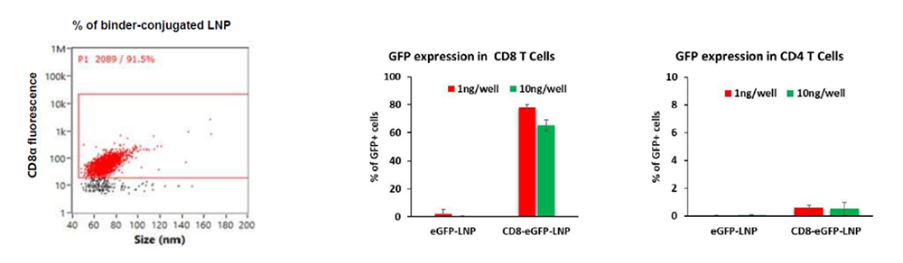
Alexa Fluor™ 488-Labeled Human CD8 alpha Protein, His Tag (Cat No. CDA-HA2H6) was used to evaluate binding and targeting of CD8-targeted lipid nanoparticles (CD8-LNPs). After, CD8-LNPs were tested to deliver eGFP to immune cells in vitro. Flow cytometry and fluorescence analysis showed high eGFP expression specifically in CD8⁺ cells, confirmed the efficient and selective targeting capability of CD8-LNPs. These results demonstrate the potential of CD8-LNPs for precise delivery of molecular cargo to CD8-expressing cell populations. (Data from Tiva Biosciences).
Product List
References
1. Srinivasan S, Zhu C, McShan AC. Structure, function, and immunomodulation of the CD8 co-receptor. Front Immunol. 2024;15:1412513. Published 2024 Aug 26. doi:10.3389/fimmu.2024.1412513
2. Glatzová D, Cebecauer M. Dual Role of CD4 in Peripheral T Lymphocytes. Front Immunol. 2019;10:618. Published 2019 Apr 2. doi:10.3389/fimmu.2019.00618
3. Ton Nu QC, Deka G, Park PH. CD8+ T cell-based immunotherapy: Promising frontier in human diseases. Biochem Pharmacol. 2025;237:116909. doi:10.1016/j.bcp.2025.116909
4. Hunter TL, Bao Y, Zhang Y, et al. In vivo CAR T cell generation to treat cancer and autoimmune disease. Science. 2025;388(6753):1311-1317. doi:10.1126/science.ads8473
 Popular ArticlesRelated RecommendationsPopular Events
 AI-Assisted Bispecific Antibody Development: From Target Pairing Prediction to Experimental Validation2026-07-31Page Views:52
 The Brain's Fountain of Youth: How Modern Neuroscience Is Decoding Healthy Brain Aging2026-07-31Page Views:22
 In Vivo CAR-T Therapy: Next-Generation CAR-T Cell Engineering and Gene Delivery Advances2026-07-29Page Views:48
 A Comprehensive Guide to Sterility Testing in Biopharmaceutical Manufacturing: From Compendial Methods to qPCR-Based Rapid Testing2026-07-27Page Views:67
 Biomarkers: Enabling Precision Assessment of Cardio-Kidney-Metabolic (CKM) Syndrome2026-07-24Page Views:97




 Related Products