



 Sep 24, 2026
 Page Views:139


Introduction
Cardiac safety remains a critical consideration in drug development. Even when a candidate demonstrates strong target activity and therapeutic efficacy, adverse cardiac effects—including QT interval prolongation, arrhythmias, myocardial injury, and impaired contractile function—may limit its progression through preclinical and clinical development. Over the past several decades, cardiac safety evaluation has evolved into a relatively mature framework, incorporating animal models such as dogs and non-human primates, hERG ion channel screening, and human-induced pluripotent stem cell-derived cardiomyocytes (hiPSC-CMs). These approaches have helped identify and eliminate many compounds with potentially cardiac liabilities. However, a fundamental question remains: how well do these non-clinical risk signals predict cardiac risk in humans?
Animal Models: Predictive Value and Translational Limitations
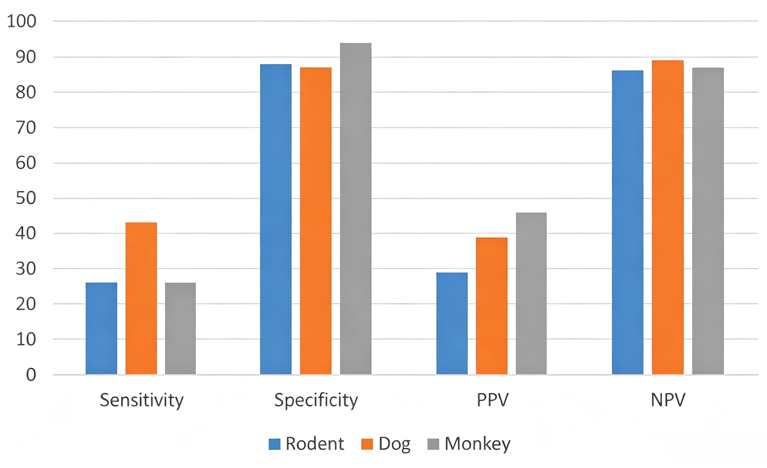
Animal models are important non-clinical evaluation tools. Compared with in vitro models, they capture cardiovascular, metabolic, and multi-organ interactions within an integrated physiological system, enabling the assessment of systemic effects that cannot be fully reproduced using isolated cells or ion channels. In an analysis of 150 drugs encompassing 221 known human toxicity findings, the combined use of rodent and non-rodent species detected approximately 71% of toxicities affecting the same target organs in humans1. Similarly, an IQ Consortium analysis of non-clinical and clinical data from 182 drugs reported that animal studies predicted clinical toxicity with a sensitivity of approximately 48%, specificity of 84%, a positive predictive value (PPV) of 43%, and a negative predictive value (NPV) of 86%2.
https://doi.org/10.1016/j.taap.2017.09.006
Consistency parameters grouped by test species
However, animal models are limited by interspecies differences in heart rate, ion channel expression and function, action potential characteristics, drug metabolism, and myocardial physiology. Therefore, cardiac safety findings from animal studies may not always translate directly to human responses3.
hERG Screening: From Ion-Channel Assessment to Integrated Cardiac Function
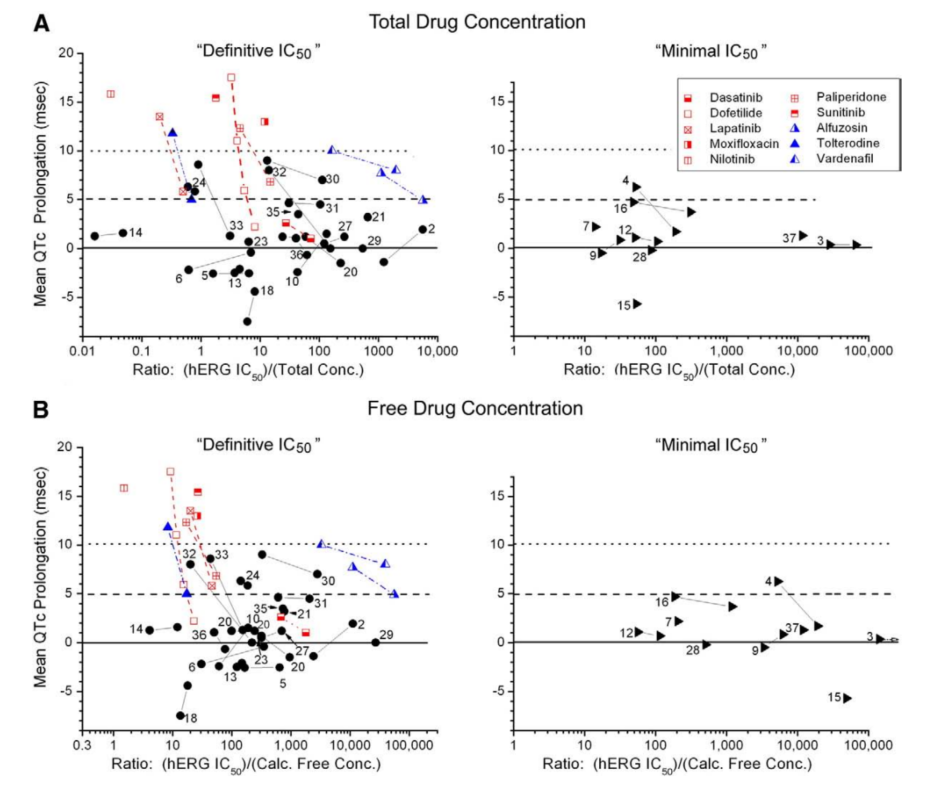
The hERG potassium channel plays an important role in cardiac repolarization, and its inhibition can increase action potential duration and the risk of QT interval prolongation, making hERG an important early screening marker in cardiac safety assessment. A retrospective analysis of 39 drugs showed that the hERG safety margin predicted clinical corrected QT interval (QTc) prolongation with a sensitivity of approximately 64%, specificity of about 88%, and an AUC of 0.724. However, cardiac repolarization is not determined by a single ion channel alone; rather, it results from the coordinated activity of multiple ion currents that collectively shape the cardiac action potential. Drug effects on multiple channels may reinforce or offset one another, leading to different electrophysiological and arrhythmic outcomes. Therefore, hERG inhibition alone is not sufficient to predict QT prolongation or the development of Torsades de Pointes (TdP).
https://doi.org/10.1016/j.pharmthera.2010.08.008
Correlation between hERG safety window and clinical QTc prolongation. The scatter plot shows that in both low safety window (<30) and high safety window (>1000) regions, there is overlap between QTc prolongation (>5 ms) and no prolongation (<5 ms), indicating that hERG blocking potency and clinical repolarization delay are not in a one-to-one correspondence. Single-channel signals are insufficient to predict pro-arrhythmic risk, necessitating a shift toward multi-level functional integration evaluation.
The Comprehensive in vitro Proarrhythmia Assay (CiPA) represents a shift in proarrhythmic risk assessment from single-endpoint testing toward a more integrated framework. Rather than relying on an individual assay, CiPA integrates multi-ion channel effects, in silico modeling, functional responses in human cardiomyocytes, and clinical data to assess proarrhythmic risk. The focus therefore moves beyond "whether an individual ion channel is inhibited" to "whether the combined effects lead to clinically meaningful changes in cardiac function."
2D Models: Human Relevance and Functional Limitations
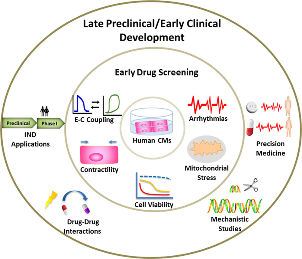
2D hiPSC-CMs have significantly improved the human relevance of cardiac safety assessment. Technologies such as microelectrode arrays (MEA), calcium imaging, and contractility assays enable direct evaluation of drug-induced changes in cardiac electrophysiology, calcium handling, and contractile function. However, the heart is a complex multicellular organ whose function depends not only on cardiomyocytes but also on their interactions with fibroblasts, endothelial cells, and other cell types, together with regulation by the extracellular matrix and local mechanical environment. Therefore, although hiPSC-CMs can effectively evaluate drug-induced electrophysiological and functional changes at the cellular level, they are less able to recapitulate tissue-level processes involving three-dimensional architecture, multicellular interactions, and longer-term remodeling, including mitochondrial dysfunction, oxidative stress, fibrotic responses, and multicellular toxicity.
https://doi.org/10.1161/CIRCRESAHA.119.315378
Key phenotypic changes that can be assessed in human cardiomyocytes (CMs) during early drug screening
Cardiac Organoids: Moving Toward More Human-Relevant Models
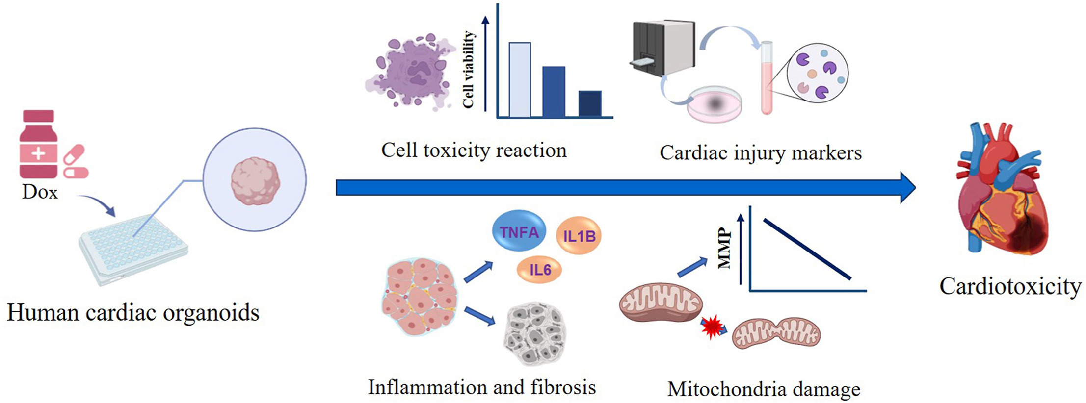
If 2D hiPSC-CMs primarily capture functional changes at the cellular level, cardiac organoids extend this assessment to the three-dimensional tissue level. The transition from 2D to 3D involves more than spatial organization, allowing cell-cell interactions, tissue structure, and functional responses to be represented in a more physiologically relevant context. Existing studies have shown that cardiac organoids can recapitulate tissue-level phenotypes such as calcium handling abnormalities, metabolic changes, fibrotic responses, and doxorubicin-induced cardiotoxicity, providing a platform for investigating complex cardiac toxicity mechanisms and strengthening preclinical-clinical translation5,6. In addition, long-term culture and repeated dosing may further expand their application from acute screening to repeated-dose and chronic toxicity studies, supporting more predictive assessment of drug-induced cardiotoxicity.
https://doi.org/10.1016/j.cbi.2023.110777
Schematic diagram of the mechanism of doxorubicin (Dox)-induced cardiotoxicity in human cardiac organoids
Conclusion: From "Human-Relevant" to "More Predictive"
From animal models to hERG and 2D hiPSC-CMs, and then to cardiac organoids, cardiac safety assessment has been developing toward models with greater human relevance and more comprehensive functional readouts. With the development of New Approach Methodologies (NAMs), regulatory agencies are increasingly emphasizing model reliability, scientific validity, and clear application contexts. Therefore, being "more human-like" is only the starting point of model development; the ability to provide stable, reproducible safety signals that are reliably associated with human risk truly determines its translational value.
Based on this concept, ACROBiosystems ready-to-use cardiac organoids (Cat. No. CIPO-HWL01) provide a standardized 3D human cardiac model for cardiac safety assessment. They require no in-house differentiation, demonstrate consistent batch-to-batch performance, and exhibit mature functional characteristics.
Case Study
Electrophysiological profiling of hiPSC-derived cardiac organoids by MEA
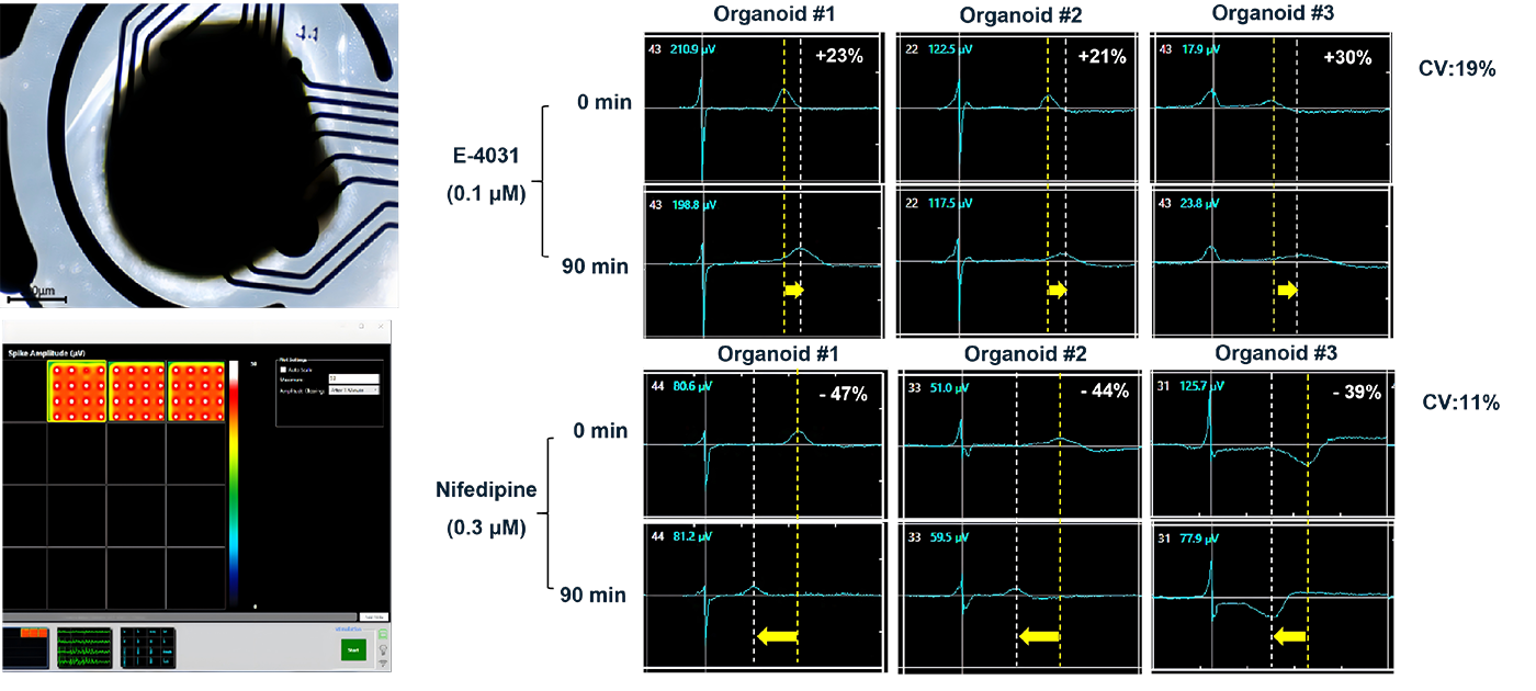
As part of functional QC, MEA recordings showed stable electrical activity across multiple electrodes in cardiac organoids. Following treatment with E-4031, a hERG/IKr potassium channel blocker, and nifedipine, an L-type calcium channel blocker, the organoids showed compound-related changes in MEA-derived electrophysiological readouts, supporting the potential use of this model for cardiac functional and safety assessment.