


 Sep 7, 2026
 Page Views:284


Introduction
Drug resistance remains one of the most persistent challenges in ADC therapy — and tumor heterogeneity is often at the root. Within a single lesion, HER2-high, HER2-low, and HER2-negative cells can coexist, while EGFR can divert HER2 into EGFR–HER2 heterodimers, reducing HER2-directed ADC internalization and efficacy. The result: incomplete drug delivery and, ultimately, treatment failure.
A study recently published in Nature proposes an unexpected solution: instead of building a single multispecific ADC upfront, the researchers delivered two separate components — an antibody and an ADC — that find each other inside the body through bioorthogonal click chemistry.
The Delivery Challenge in ADC Development
Approximately a dozen ADCs have received FDA approval. However, their activity can still be limited by two closely related challenges: tumor heterogeneity and treatment resistance.
Challenge 1: Tumor Heterogeneity
Antigen expression can vary substantially within the same tumor. HER2-high, HER2-low, and HER2-negative cell populations may coexist, limiting the delivery of a HER2-directed ADC to cells with low or absent target expression. These insufficiently targeted populations may survive and contribute to disease progression or recurrence.
Challenge 2: Treatment Resistance
Tumors can become less responsive to ADC treatment through antigen downregulation, altered receptor internalization, or activation of compensatory signaling pathways.
The Nature study examined EGFR upregulation as one such mechanism. EGFR can promote EGFR–HER2 heterodimer formation, reducing the internalization and activity of HER2-directed ADCs.
Both challenges reflect a common limitation: conventional ADCs typically depend on a single antigen for tumor localization and cellular uptake. The antibody–ADC click strategy provides a complementary approach by connecting an antibody and an ADC through bioorthogonal chemistry following sequential administration.
How Antibody–ADC Click Works
The platform uses the inverse electron-demand Diels–Alder reaction between trans-cyclooctene (TCO) and tetrazine (Tz). This rapid bioorthogonal reaction proceeds without a catalyst under physiological conditions.
Administration of the targeting antibody. Panitumumab, an EGFR-targeting antibody, was modified with TCO and administered first. The antibody distributed systemically and bound to EGFR-expressing tumor cells.
Administration of the ADC. Twenty-four hours later, a tetrazine-modified HER2-directed ADC—either trastuzumab deruxtecan (T-DXd) or trastuzumab emtansine (T-DM1)—was administered.
The complementary TCO and tetrazine groups formed a covalent linkage following systemic delivery, generating antibody–ADC click constructs capable of engaging both receptors.
The study found that ligation could occur both in circulation and after tumor accumulation. The observed effects therefore probably reflect a combination of bioorthogonal ligation, tumor targeting, receptor engagement, internalization, and changes in distribution.
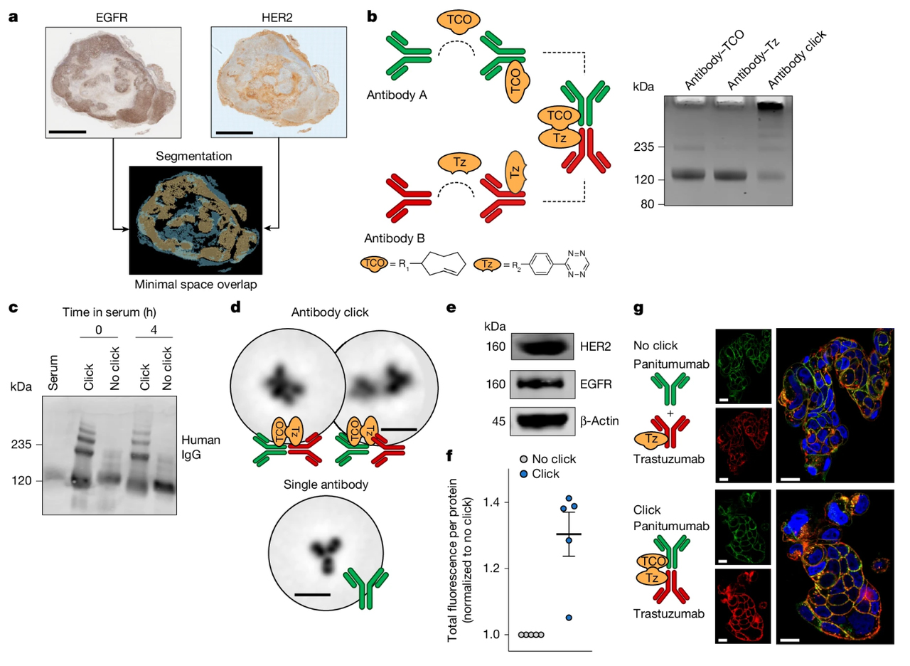
Figure 1. Antibody click platform for in vivo ligation of antibody–ADC constructs via bioorthogonal chemistry.
Source: (doi: 10.1038/s41586-026-10789-w)
Enhanced Internalization Across HER2 Expression Levels
Confocal microscopy and live-cell imaging showed that clicked constructs increased internalization in multiple cancer cell lines compared with non-clicked antibody combinations. The resulting complexes were also transported to lysosomes, an important step in intracellular payload delivery.
In this study, EGFR engagement provided an additional route for the uptake of HER2-directed antibodies and ADCs in cells with limited HER2 expression. However, the experiments could not definitively separate the contributions of receptor-mediated uptake, antibody clustering, and pharmacokinetic changes caused by antibody ligation.
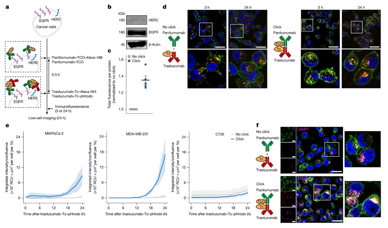
Figure 2. Antibody click enhances internalization in HER2− cancer cell lines.
Source: (doi: 10.1038/s41586-026-10789-w)
Preclinical Evidence: Distribution, Uptake, and Efficacy
Limited Spatial Overlap Between HER2 and EGFR
The researchers first used automated image alignment and segmentation to analyze HER2 and EGFR expression in tumor biospecimens. Regions with high HER2 and EGFR expression showed limited spatial overlap, with reported Dice similarity coefficients of 0.12 and 0.26, where 0 represents no overlap and 1 represents complete overlap.
The study then used a bilateral tumor model to mimic intertumoral heterogeneity. Individual mice carried two xenografts with distinct receptor profiles:
- HER2-positive, EGFR-low NCI-N87 tumors
- HER2-ultralow, EGFR-high A431 tumors
The model allowed the researchers to assess whether targeting both receptors could improve ADC distribution across tumor regions that would not be uniformly accessible through HER2 alone.
A 3.2-Fold Increase in Uptake in HER2-Ultralow Tumors
At 24 hours, uptake in HER2-ultralow A431 tumors increased from approximately 3.75% injected dose per gram (%ID/g) with trastuzumab–tetrazine alone to approximately 11.90%ID/g with antibody click—a 3.2-fold increase.
In HER2-positive NCI-N87 tumors, uptake was similar between the two groups:
| Tumor model | Trastuzumab–Tz alone | Antibody click | Difference |
|---|---|---|---|
| HER2-ultralow A431 | ~3.75%ID/g | ~11.90%ID/g | 3.2-fold increase |
| HER2-positive NCI-N87 | ~28.90%ID/g | ~31.63%ID/g | Comparable |
Control experiments using radiolabeled non-targeting IgG–tetrazine showed that click ligation could retarget a second antibody toward the antigen recognized by the first antibody. However, simultaneous targeting of HER2 and EGFR produced greater uptake of the second, drug-bearing antibody than a non-targeted carrier.
Antitumor Activity in Heterogeneous Models
In the bilateral NCI-N87/A431 model, mice received a single intravenous dose of panitumumab or panitumumab–TCO at 5 mg/kg, followed 24 hours later by T-DXd or T-DXd–tetrazine.
The T-DXd click group showed slower or halted tumor progression compared with vehicle and no-click controls in both tumor types. All mice in the T-DXd click group showed tumor regression (fold change below baseline) in HER2-positive NCI-N87 tumors.
The researchers also evaluated breast and pancreatic cancer xenograft models with low or undetectable HER2 expression. These tumors showed minimal responses to HER2-directed ADC monotherapy or non-clicked panitumumab-plus-ADC combinations. In contrast, the corresponding antibody–ADC click treatments reduced tumor growth and prolonged survival.
These findings demonstrate activity in the tested preclinical models but do not establish safety or efficacy in humans.
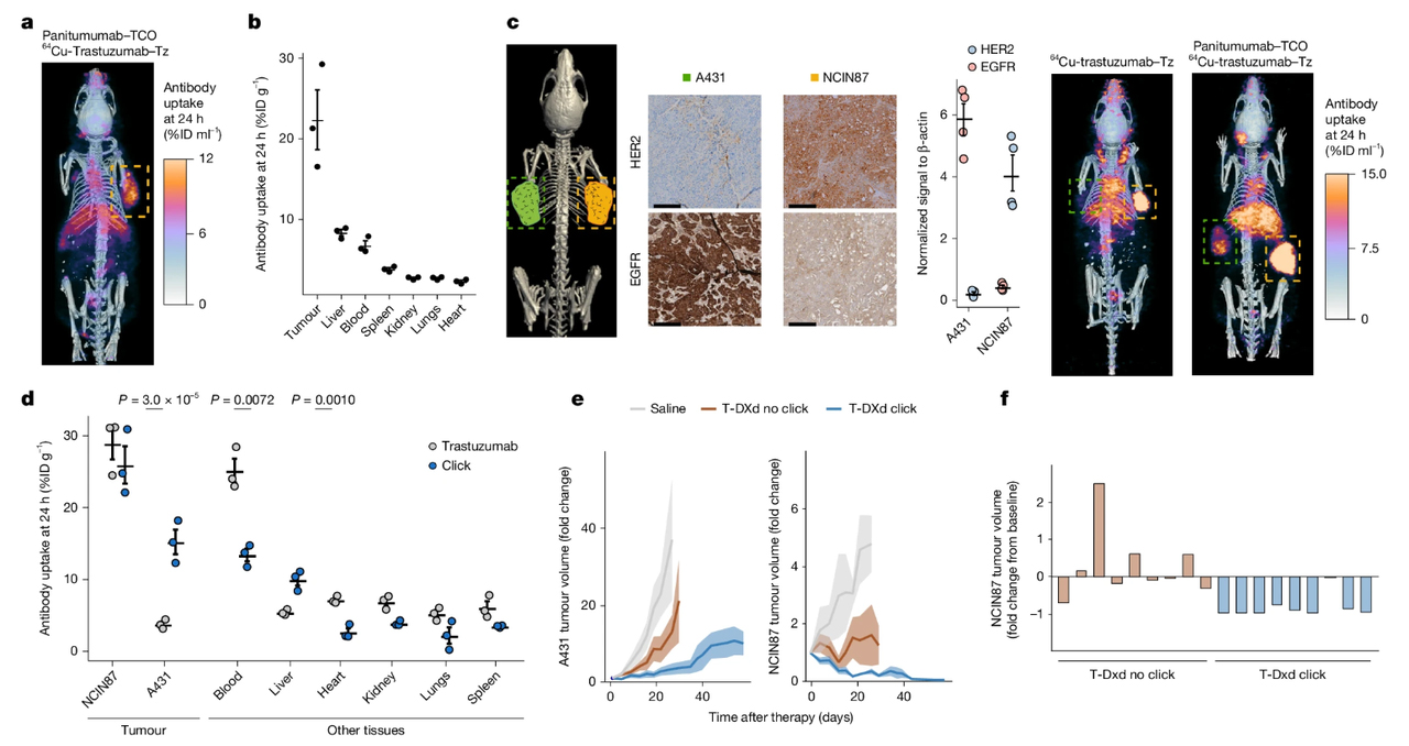
Figure 3. Antibody-ADC click enhances tumour uptake and therapeutic efficacy.
Source: (doi: 10.1038/s41586-026-10789-w)
Activity in T-DXd-Nonresponsive and Resistant Models
The study further examined whether the platform could improve responses in tumors that were unresponsive or resistant to HER2-directed treatment.
Mice with HER2-positive tumors were initially treated with T-DXd and classified as responders or nonresponders. Tumors from the nonresponding animals had 2.1-fold higher total EGFR protein levels than those from responding animals.
After the nonresponding mice were switched to panitumumab–TCO plus T-DXd–tetrazine, tumor growth was reduced in 5 of 9 animals.
The researchers also evaluated a trastuzumab-resistant BT474 model. Six of 10 mice were classified as nonresponders to T-DXd. After these 6 animals were switched to pertuzumab–TCO plus T-DXd–tetrazine, tumor suppression was observed in 5 of 6 non-responder mice.
These results support further investigation of antibody–ADC click in refractory tumor models. They should not be interpreted as evidence that the approach can reverse ADC resistance in patients.
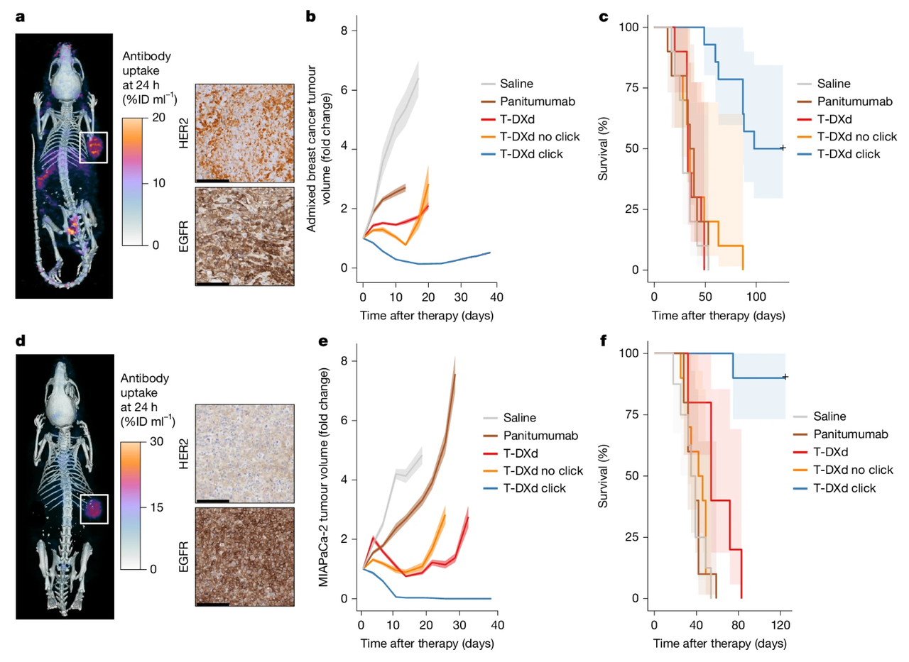
Figure 4. Antibody-ADC click overcomes tumour resistance to T-DXd.
Source: (doi: 10.1038/s41586-026-10789-w)
A Modular Antibody–ADC Platform
The study demonstrated flexibility at several levels.
1. Antibody Pairing
The researchers evaluated multiple combinations, including panitumumab plus trastuzumab and pertuzumab plus trastuzumab. The approach supported antibodies recognizing different receptors as well as antibodies binding different epitopes of the same receptor.
2. ADC Selection
Both T-DXd and T-DM1 were evaluated as HER2-directed ADC components, demonstrating that the click strategy was not limited to a single ADC in the reported models.
The conjugates carried approximately 3–12 TCO or tetrazine groups per antibody. Under the reported experimental conditions, these modifications did not measurably disrupt cell binding.
3. Potential Expansion
HER2 and EGFR served as the proof-of-concept receptor pair. The modular design may provide a basis for evaluating other receptors, including HER3, TROP2, c-Met, and Claudin 18.2, as well as other payload classes.
These possibilities remain future development directions rather than combinations validated in the reported study. Each new antibody, ADC, target pair, and conjugation format would require independent characterization.
The authors also note that further development for clinical translation will require continued evaluation and optimization of pharmacokinetics, linker or spacer design, construct size and valency, and payload-related safety.
Conclusion
The study by Simó and colleagues presents a different way to think about ADC design. Rather than remaining a single, fixed construct, an ADC may function as one component of a modular system assembled through bioorthogonal ligation following sequential administration.
In the reported preclinical models, this strategy improved the delivery and activity of HER2-directed ADCs across heterogeneous HER2 expression levels and in tumors that were unresponsive or resistant to conventional HER2-directed treatment.
However, the platform remains at the preclinical stage. Further optimization and evaluation will be required before its translational potential can be determined.
Supporting ADC Research with Fit-for-Purpose Reagents
The antibody–ADC click study highlights the importance of evaluating target expression, antibody internalization, intracellular trafficking, and pharmacokinetics when developing new ADC formats.
ACROBiosystems provides reagents and assay solutions for ADC discovery and characterization.
pHintra™ Internalization Detection Solutions
pHintra™ Internalization Detection Reagent binds the Fc region of human IgG, enabling internalization assessment. Labeling can be completed in approximately 10 minutes, followed by detection using flow cytometry or cell imaging.
pHintra™ Antibody Labeling Kit supports covalent labeling of antibodies with a pH-sensitive fluorescent dye. It can be used with antibodies from different species and isotypes for flow cytometry and imaging applications.
ADC Target Proteins and Functional Cell Lines
Recombinant proteins covering 90+ ADC-related targets are available with multiple species and tag options.
Target overexpression cell lines can support cell-based binding, internalization, and functional assay development.
ADC Pharmacokinetic Assay Reagents and Solutions
ACROBiosystems offers reagents and assay solutions supporting conjugated antibody quantification, ADC pharmacokinetic assay development, and immunogenicity assessment.
These products are intended to support researchers in establishing and validating their own ADC experiments. Assay formats and conditions should be selected according to the antibody format, target biology, cell model, and specific research question.

_ Clinical Progress and Emerging Therapeutic Strategies in Solid Tumors.jpg)